Viral
Dina Rickman
Mar 03, 2015
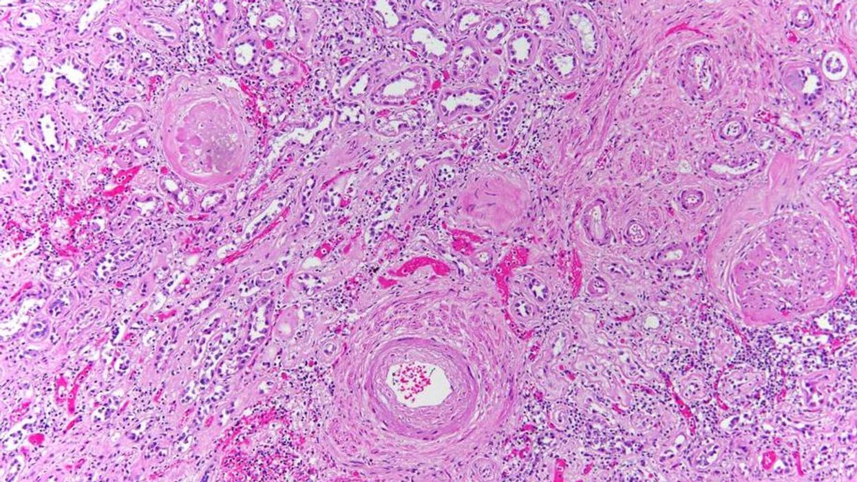
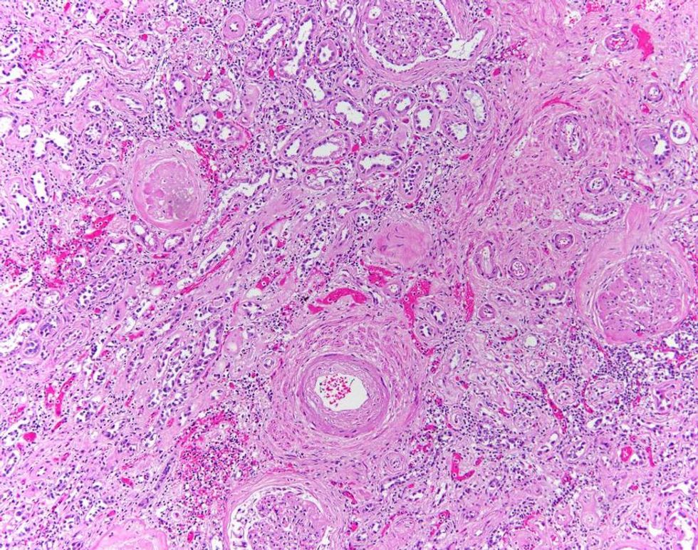
I really like looking at kidneys under the microscope. There’s part of the kidney that would make incredible wallpaper.
So says pathologist Dr Marianne Hamel of her favourite body part to examine. This is a woman who knows what she is talking about: her exhibition of microscopic art Death Under Glass opens in London from Tuesday evening after generating worldwide press attention and plaudits in Philadelphia.
The kidney
Dr Hamel developed the exhibition after remarking to forensic photographer Nikki Johnson the images she saw under the microscope while performing autopsies were "beautiful". In response, Johnson suggested they work together to share them with the public.
The aim behind the show is twofold: firstly to showcase the importance of histological analysis (“one of the bedrocks of pathology”) and also so the world can see what the body looks like close up.
Small bowel
“Previously, if you wanted to see what I see you had to sit with me with a double headed microscope and I'd take you through it,” Dr Hamel told i100.co.uk.
The majority of the images are representative of what Dr Hamel sees when performing autopsies. The colours are diagnostic colours which are added to slides to help define human tissue.
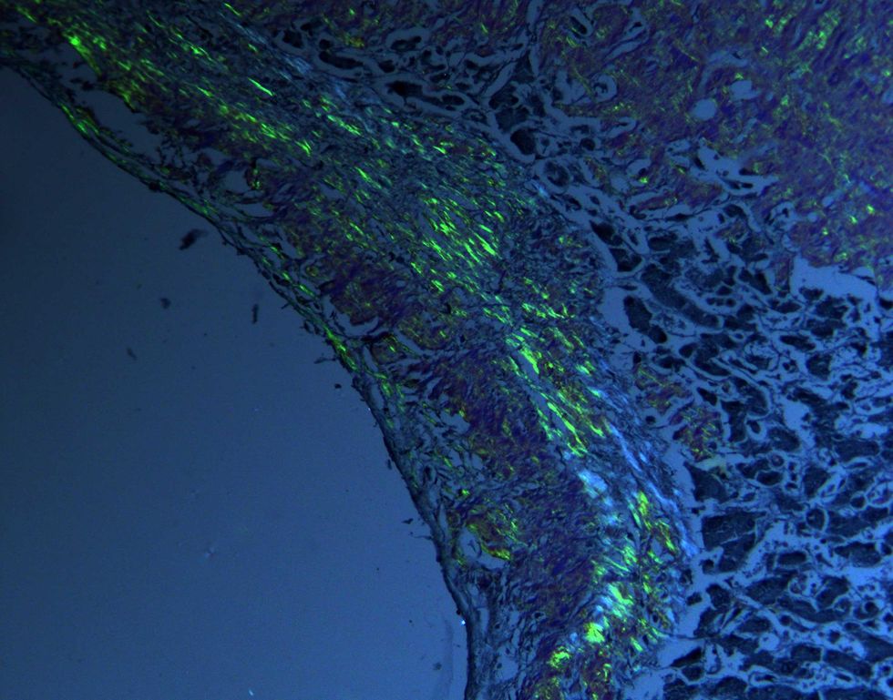
Birefringence
“Histology is underrated as a design element,” she says. “The public’s reaction to it has largely been very positive. I don’t think they find it ghoulish, I think they see it as very beautiful.”
Gunshot wound
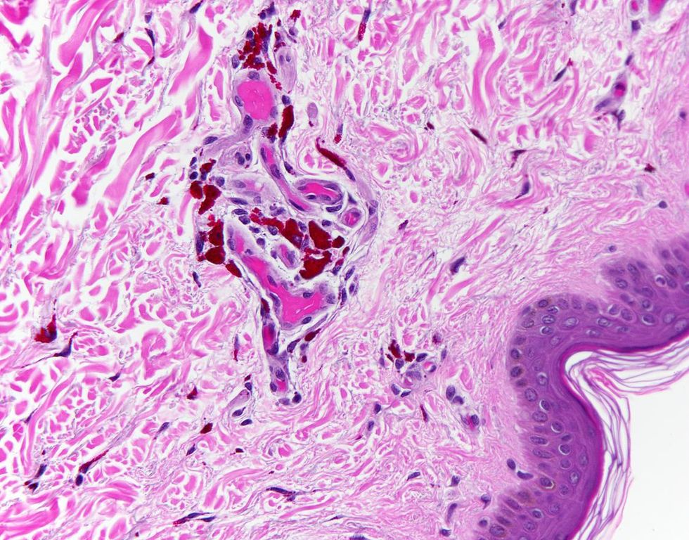
Tattoo
‘Death Under Glass’ launches on Wednesday 3 March at Barts Pathology Museum. For more details click here.
Top 100
The Conversation (0)
